Category: Blogs

  • Know about Joint Replacement Surgery? Best Orthopedic Surgeons

    Know about Joint Replacement Surgery? Best Orthopedic Surgeons

    What is Joint Replacement Surgery? Get Recovery Tips from Best Orthopedic Surgeons

    joint replacement surgery

    Joint pain is one of the leading causes of disability worldwide, affecting millions of people each year. For those suffering from chronic joint pain due to arthritis, injury, or other degenerative conditions, joint replacement surgery can offer a new lease on life. This comprehensive guide will explore everything you need to know about joint replacement surgery, including the different types of procedures, what to expect before and after surgery, and expert tips from leading orthopedic surgeons to ensure a smooth and successful recovery.

    Understanding Joint Replacement Surgery

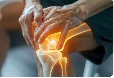
    Joint replacement surgery, also known as arthroplasty, is a procedure in which a damaged joint is removed and replaced with a prosthetic implant. These implants are designed to mimic the function of natural joints, restoring mobility and alleviating pain. This type of surgery is most commonly performed on the hips and knees, but it can also be done on shoulders, elbows, ankles, and fingers.

    Why Joint Replacement Surgery is Performed

    Joint replacement surgery is typically recommended for individuals who have severe joint damage that limits daily activities and does not respond to conservative treatments such as physical therapy, medication, or injections. Common causes include: Osteoarthritis, Rheumatoid arthritis, Post-traumatic arthritis, Congenital joint disorders, Avascular necrosis, Joint deformities, Severe fractures & more.

    Types of Joint Replacement Surgeries

    Total Joint Replacement: In total joint replacement, both sides of a joint are replaced with prosthetic components. For example, in a total knee replacement, the ends of the femur and tibia are replaced with metal and plastic components.

    Partial Joint Replacement: Partial joint replacement involves replacing only the damaged part of the joint. This is often used when damage is limited to one compartment, as in partial knee replacements.

    Revision Joint Replacement: Sometimes, a joint replacement may need to be redone due to wear, infection, or implant failure. This is known as revision surgery and is more complex than primary replacement.

    Minimally Invasive Joint Replacement: This technique uses smaller incisions and specialized instruments to reduce tissue damage and speed up recovery.

    Robotic-Assisted Joint Replacement: The rise of robotic-assisted surgery has marked a major advancement in joint replacement. This technique uses a computer-assisted robotic arm that helps the surgeon plan and execute the procedure with extremely high precision.

    Common Conditions you to Needs Joint Replacement Surgery

    Several underlying diseases and conditions contribute to joint damage that may ultimately require surgical intervention.

    a) Osteoarthritis (OA)

    The most common indication for joint replacement surgery, OA is a degenerative condition where the cartilage that cushions the joint surfaces wears down over time. OA is especially prevalent in knees, hips, and shoulders. When joint space narrows significantly and bone rubs against bone, surgery becomes necessary to restore comfort and mobility.

    b) Rheumatoid Arthritis (RA)

    This autoimmune condition causes chronic inflammation of the joints, leading to cartilage destruction, bone erosion, and deformity. While newer biologic drugs can control inflammation, longstanding RA may still result in the need for joint replacement—especially in hands, wrists, shoulders, and knees.

    c) Post-Traumatic Arthritis

    A fracture or injury that damages the cartilage or joint structure can accelerate arthritis. Even after the bone heals, the joint may never function properly again. Recurrent instability, stiffness, or pain after injury can prompt consideration for surgical repair or replacement.

    d) Avascular Necrosis (AVN)

    Avascular necrosis occurs when the blood supply to a joint bone (typically the femoral head) is disrupted, causing the bone to collapse and the joint to fail. Early AVN can be treated conservatively, but advanced cases often require hip or shoulder replacement.

    e) Congenital or Structural Abnormalities

    Some individuals are born with malformed joints (e.g., hip dysplasia) or acquire misalignments over time (e.g., leg length discrepancies). These can cause abnormal wear patterns, leading to premature joint deterioration that necessitates surgical correction.

    Factors That Affect Surgical Eligibility

    Even if someone qualifies for surgery based on joint damage and symptoms, other health and lifestyle factors can influence whether it’s safe or advisable to proceed.

    a) Age

    There is no strict age limit, but:

    • Patients under 50 may be discouraged from getting joint replacements unless necessary, due to concerns about implant longevity and future revisions.

    • Older patients over 80 can undergo surgery safely, but overall fitness and comorbidities must be considered.

    b) Body Weight

    Obesity (especially BMI > 40) increases the risk of complications like infection, blood clots, and implant failure. Many surgeons encourage patients to lose weight before surgery.

    c) Chronic Illnesses

    Uncontrolled diabetes, heart disease, or respiratory conditions may delay surgery until stabilized. A thorough pre-op medical evaluation ensures the patient is fit for anesthesia and recovery.

    d) Smoking and Alcohol

    Smoking impairs wound healing and increases infection risk. Surgeons often require patients to stop smoking at least 4–6 weeks before and after surgery.

    e) Mental Health and Support System

    A strong support system, good mental health, and compliance with post-op instructions are essential. Depression, anxiety, or poor social support can negatively affect outcomes.

    Ready to Take the First Step Toward a Pain-Free Life?

    Don’t let joint pain limit your movement or quality of life. Whether you’re considering joint replacement surgery or just need expert guidance on your symptoms, our experienced orthopedic specialists are here to help.

    📞 Book Your Consultation Today

    joint replacement surgery

    FAQs About Kidney Stones Symptoms

    Joint replacement surgery is a medical procedure where a damaged or diseased joint is replaced with an artificial prosthesis made of metal, plastic, or ceramic. It’s typically needed when joint pain, stiffness, or deformity caused by arthritis, injury, or degenerative disease severely affects a person’s quality of life and conservative treatments like medications or therapy no longer help.

    The most common types include:

    • Knee Replacement (TKR or UKR) – for severe osteoarthritis or injury

    • Hip Replacement (THR) – for hip fractures or arthritis

    • Shoulder Replacement – for rotator cuff damage or arthritis

    • Ankle and Elbow Replacement – less common, used for trauma or rheumatoid arthritis

    Each procedure is tailored to the specific joint and the severity of the condition.

    Most joint replacement surgeries last between 1 to 3 hours, depending on the joint being replaced and the complexity of the case. This is followed by a few hours in a recovery room under observation.

    Pain during surgery is managed through general anesthesia or spinal/epidural blocks, so patients don’t feel pain during the procedure. Post-operative pain is common but manageable with medications and gradually reduces as healing progresses.

    Recovery time varies:

    • Knee and Hip Replacement: 6–12 weeks for basic activities, up to 6–12 months for full recovery.

    • Shoulder Replacement: 3–6 months.

    • Ankle/Elbow Replacement: 3–6 months.

    Physical therapy, age, general health, and adherence to post-op instructions all affect recovery time.

    While most surgeries are successful, potential complications include:

    • Infection

    • Blood clots (DVT or pulmonary embolism)

    • Implant loosening or dislocation

    • Nerve or vessel injury

    • Joint stiffness

    • Allergic reaction to implant materials

    Most complications are rare and can be prevented or managed with early detection and proper care.

    Related Post

    Book Appointment

  • Understanding Kidney Stone Symptoms: Causes, Pain Relief

    Understanding Kidney Stone Symptoms: Causes, Pain Relief

    Understanding Kidney Stone Symptoms: Causes, Pain Relief & Diet Tips to Prevent Them

    Understanding kidney stone symptoms

    Kidney stones are a painful and increasingly common health issue affecting people of all ages. Whether you’ve had them once or fear their onset due to family history or lifestyle, understanding the kidney stone symptoms, causes, and how to prevent them through an effective diet plan for kidney stone can help you stay ahead of this painful condition. Let’s walk through everything you need to know in a detailed yet conversational way.

    The real pain of kidney stones doesn’t start until they move. While they’re in the kidney, you might not feel a thing. But once a stone shifts into the ureter—the narrow tube that connects your kidney to your bladder—it can cause intense pain. The ureter is not designed to stretch like the bladder, so when a stone tries to push through, it causes cramping, inflammation, and sometimes complete blockage of urine flow. This pressure buildup is what makes the pain almost unbearable and often sends people rushing to the emergency room.

    Common Kidney Stone Symptoms

    Pain in the Lower Back or Side: The hallmark of kidney stones is a sudden, sharp pain in the side or lower back, usually below the ribs. This pain might radiate to the lower abdomen or groin and can come in waves, increasing and decreasing in intensity. It’s often described as one of the most intense types of pain a person can experience. If you’re doubling over and can’t find a comfortable position, a kidney stone could be the culprit.

    Painful Urination and Frequent Urge: Passing a kidney stone can make urinating excruciating. You may feel a burning sensation or an intense urgency, even if you’re unable to produce much urine. This is because the stone irritates the lining of the urinary tract, making the bladder and urethra hypersensitive.

    Blood in Urine: Seeing pink, red, or brown urine can be alarming, but it’s a common symptom of kidney stones. The stone can scrape the walls of the urinary tract, causing bleeding. Sometimes, the blood is only visible under a microscope, but in other cases, it can noticeably discolor your urine.

    Nausea and Vomiting:  Due to the shared nerve pathways between the kidneys and the gastrointestinal tract, severe kidney stone pain often comes with nausea and vomiting. When your body senses something is wrong, especially in your urinary system, it may trigger your digestive system to shut down or react adversely.

    Foul-Smelling or Cloudy Urine: Another common symptom is urine that appears cloudy or has a strong, unpleasant odor. This could indicate a urinary tract infection (UTI), which sometimes develops when a kidney stone causes a blockage and bacteria begin to multiply.

    Fever and Chills: If you develop a fever or chills along with the other symptoms, it may mean a kidney infection is setting in—a dangerous and potentially life-threatening condition. This requires immediate medical attention.

    Causes of Kidney Stones

    Dehydration: One of the most common and preventable causes of kidney stones is simply not drinking enough water. When you’re dehydrated, your urine becomes more concentrated, meaning there’s less fluid to dissolve the minerals that can turn into stones. This creates a perfect storm for stone formation. Make it a habit to drink water throughout the day, especially if you sweat a lot or live in a hot climate.

    Excessive Salt or Animal Protein Intake: Too much salt increases the amount of calcium your kidneys must filter, which raises the chances of forming calcium-based stones. Similarly, eating too much red meat, eggs, or seafood can raise uric acid levels, another contributor to stone formation. Moderation and balance are key when it comes to salt and protein.

    Genetic Predisposition: If you have a family history of kidney stones, your chances of developing them are significantly higher. Genetics can influence how your body processes certain minerals and fluids. While you can’t change your genes, knowing your risk can help you make better lifestyle choices.

    Certain Medical Conditions: Conditions like urinary tract infections (UTIs), obesity, and hyperparathyroidism can increase your risk of stones. People with inflammatory bowel disease (IBD) or those who’ve had gastric bypass surgery may also be at greater risk due to changes in calcium and oxalate absorption.

    Medications and Supplements: Ironically, certain supplements and medications can contribute to kidney stones. Calcium supplements (especially when taken in excess), vitamin D, antacids, and even frequent use of laxatives can increase your risk. Always consult your doctor before starting any new supplement, especially if you have a history of stones.

    Types of Kidney Stones

    Calcium Oxalate Stones: These are the most common and are caused when calcium combines with oxalate in your urine. They’re often linked to dehydration, high oxalate foods, and low calcium diets. Foods high in oxalate include spinach, beets, nuts, and chocolate.

    Uric Acid Stones: These form when your urine is too acidic. They’re common in people who consume a lot of red meat or are undergoing chemotherapy. Uric acid stones are also associated with gout and diabetes.

    Struvite Stones: Typically found in women with recurrent UTIs, struvite stones can grow rapidly and become quite large. They’re made up of magnesium, ammonium, and phosphate and often require medical removal due to their size.

    Cystine Stones: These are rare and develop in people with a genetic condition called cystinuria, where the kidneys excrete too much of the amino acid cystine. Managing these usually involves both medication and lifestyle changes.

    Pain Relief Options for Kidney Stones

    Over-the-Counter Pain Relievers: Mild stones can often be managed at home with over-the-counter medications like ibuprofen, naproxen, or acetaminophen. These help reduce inflammation and relieve pain while waiting for the stone to pass.

    Prescription Medications: Doctors may prescribe alpha-blockers like tamsulosin, which help relax the muscles in your ureter, allowing the stone to pass more easily and with less pain. These are typically used when the stone is small enough to pass naturally.

    Hydration Therapy: One of the first things your doctor will recommend is drinking plenty of fluids to flush out the stone. Water is best, but in some cases, doctors may suggest beverages high in citrate like lemon water, which can help dissolve the stone.

    Medical Procedures: If the stone is too large or causing complications, procedures like shock wave lithotripsy (breaking up the stone with sound waves), ureteroscopy (using a scope to remove it), or even surgery may be required.

    Sample Diet Plan for Kidney Stone Prevention

    Morning Routine: Start your day with a glass of warm lemon water to boost hydration and citrate levels. Avoid caffeinated drinks first thing in the morning, as they can lead to dehydration.

    Breakfast Options: Choose a bowl of oatmeal with almond milk, topped with berries. Whole-grain toast with low-fat cheese or a boiled egg makes a balanced start.

    Lunch Ideas: Grilled chicken with brown rice and steamed vegetables like carrots and zucchini works great. Add a side salad dressed in olive oil and lemon juice.

    Evening Snacks: A banana or a handful of watermelon cubes work well. Avoid salty snacks or anything deep-fried.

    Dinner Suggestions: Opt for baked fish or tofu, paired with quinoa and sautéed green beans. Avoid heavy sauces or gravies that are often high in salt.

    Bedtime Tips: Sip on warm water or chamomile tea before bed. Don’t forget to urinate before sleeping to avoid overnight urine concentration.

    Consult Our Expert Team Today

    If you’ve experienced any symptoms mentioned above or simply want to understand your risk factors, don’t wait. Book a consultation with our kidney health specialists today to receive a personalized assessment, diet recommendations, and long-term prevention strategies.

    kidney stone symptoms

    FAQs About Kidney Stones Symptoms

    Several factors increase your risk:

    Dehydration: Less water = more concentrated urine = higher stone risk.

    Diet: High in salt, oxalates, and animal proteins.

    Family history: Genetic predisposition increases likelihood.

    Obesity or digestive disorders: These can affect how your body processes calcium and oxalate.

    Medical conditions: Such as hyperparathyroidism, gout, and UTIs.

    Understanding and managing these risks can greatly reduce your chances of forming stones.

    Yes, especially if left untreated. Potential complications include:

    Kidney infection (pyelonephritis)

    Obstruction of urine flow, leading to swelling (hydronephrosis)

    Permanent kidney damage in severe or recurrent cases

    This is why prompt diagnosis and treatment are crucial.

    Milk and other calcium-rich foods are beneficial. They bind oxalates in your gut, reducing stone risk. But avoid calcium supplements unless advised by a doctor.

    It varies:

    Small stones (<5mm): Often pass within a few days to weeks with hydration and medication.

    Larger stones: May take longer or require medical intervention.
    The process can be painful, but proper care and pain management can ease the journey.

    Related Post

    Book Appointment

  • Abdominoplasty (Tummy Tuck) – Procedure, Benefits, & Risks

    Abdominoplasty (Tummy Tuck) – Procedure, Benefits, & Risks

    Abdominoplasty (Tummy Tuck) – Procedure, Benefits, & Risks

    What is Abdominoplasty?

    Abdominoplasty, commonly called as “tummy tuck“, is an operation to remove excess fat and skin from the abdomen. The surgeon may also tighten the tummy muscles. The aim is to produce a tighter, flatter stomach and it may partially reduce the appearance of stretch marks. Why is it required? If you have any or some of the underlying conditions, you may want to consider this option

    • Abdominal skin that is excess or sagging

    • A protruding abdomen that is out of proportion to the rest of your body

    • Abdominal muscles that have been separated and weakened

    • Excess concentrated fat tissues around your abdomen, detected by abdomen ultrasound.

    Any plans of pregnancy in future or of losing a significant amount of weight must be discussed with your plastic surgeon. Any resultant scarring from previous abdominal surgery may limit the results of your Abdominoplasty, A tummy tuck is not a treatment for weight control and is not a substitute for regular physical activity and a healthy balanced diet.

    What are the Alternatives?

    (Please note that some alternative treatments may not be available or suitable to everyone.) The purpose of tummy tuck plastic surgery is to change your body shape, not to lose weight. If fat is localized in the abdomen, but there is no excessive skin, then liposuction is an alternative to tummy tuck. Liposuction and tummy tuck are not usually done at the same time, because suction of the skin during liposuction may damage the skin that is being stretched and repositioned.

    Obese patients, or those who have large amounts of loose excess skin, may undergo a modification of the tummy tuck procedure called panniculectorny. For less severe conditions, you may want to consider the less-invasive mini tummy tuck

    How to Get Prepared for the Surgery?

    Your surgeon will give you specific instructions on how to prepare for surgery. including guidelines on

    Diet & eating habits

    Overexposure to the sun before surgery, especially to your abdomen

    Smoking & drinking

    If you smoke, plan to quit at least one to two weeks before your surgery and not to resume for at least two weeks after your surgery

    Taking or avoiding certain vitamins, and medications

    Aspirin and Vitamin E containing medications must be avoided at least 7days prior to your surgery.Specific instructions on this will be given by your surgeon.

    Carefully following these instructions will help your surgery go more smoothly. However, if you develop a cold or infection of any kind, your surgery will probably be postponed. Whether your surgery is done on an outpatient or inpatient basis, you should arrange for someone to drive you home after your surgery, and to help you out for a day or two after you leave the hospital.

    What Type of Anesthesia can be Used?

    Your doctor may select general anesthesia, so you’ll sleep throughout the operation. Other surgeons use local anesthesia, combined with a sedative to make you drowsy.

    What Happens During the Procedure?

    A tummy tuck usually begins with a long incision across the abdomen, from hip bone to hip bone. A second incision is made to free the navel from the abdominal tissue. The skin is separated from the abdominal wall, up to the ribs, exposing the vertical abdominal muscles (rectus muscles). The rectus muscles are then stitched into a new position, tightening the muscles and reducing the waistline. After the muscles have been repositioned and tightened, the excess skin is stretched and removed. The remaining skin is rewrapped over the abdominal area and sutured in place. The hole created by the incision around the belly button is normally removed when the excess skin is cut away, and a new hole is formed to place the navel proportionally.

    What Happens After the Procedure?

    For the first few days

    Abdomen will probably be swollen

    You’re likely to feel some pain and discomfort which can be controlled by medication

    Depending on the extent of the surgery, you may be released within a few hours or you may have to remain hospitalized for two to three days. Your doctor will give you instructions for

    Showering

    Changing your dressings

    All though you may find it difficult to stand straight in the beginning but make sure you start walking as soon as possible. Surface stitches will be removed in seven to ten days. The dressing on your incision may be replaced by a support garment.

    What Are the Benefits of this Procedure?

    Abdominoplasty, whether partial or complete, produces excellent results for patients with weakened abdominal muscles or excess skin. And in most cases, the results are long lasting, if you follow a balanced diet and exercise regularly.

    When to Contact your Doctor?

    After the surgery contact your doctor if

    You develop a fever over 100°F (37.8°C).

    You have drainage from your incision, or the incision separates.

    You become dizzy or faint.

    You have nausea and vomiting. You have chest pain.

    You become short of breath.

    What are the Expectations from the Surgery?

    Expectations from the surgery

    • Abdominoplasty can enhance your appearance and your self-confidence, but it won’t necessarily change your looks to match your ideal, or cause other people to treat you differently

    General Risks of having an Operation:

    These have been mentioned in the “Anesthesia Consent Form.” Please discuss this with your Anesthetist before signing the Anesthesia Consent Form.

    What are the Risks of the Procedure?

    While majority of patients have an uneventful surgery and recovery, few cases may be associated with complications. These are seen infrequently and not all the ones listed below are applicable to one individual. However, it is important that you are aware of the complications/risks that may arise out of this procedure which are as below:

    Note: The listed risks and complications are not all inclusive

    • A long, horizontal (across) scar, which is noticeable but usually fades in time Sometimes the scars can be red, thick and itchy

    • The pubic hairline may be pulled out

    • A scar like an upside-down T, or occasionally two scars, a horizontal (crosswise) one and a transverse (up and down) scar

    • Necrosis (dying) of remaining skin due to poor circulation where there is scar tissue

    • The navel may not be central Loss of the navel, the scar may provide a reasonable substitute

    • Muscle soreness immediately after operation

    • Numbness of skin pulled down below the navel for up to a year or more

    • Increased risk of clots in the leg for patients who are on the oral contraceptive pill. Part of this clot may break off and go to the lungs, which can be fatal

    • Collection of fluid under the skin above the horizontal scar

    • Marked increase of failure of the procedure due to effects of smoking on quality of blood supply to the skin of the lower part of the abdominal wall

    Tags :
    Abdominoplasty, Blogs
    Post Related

    Book Appointment

  • What Is Knock Knee: Causes, Symptoms, and Treatment Options

    What Is Knock Knee: Causes, Symptoms, and Treatment Options

    What Is Knock Knee: Causes, Symptoms, and Treatment Options

    What Is Knock Knee: Causes, Symptoms, and Treatment Options

    Knock knee, also known as genu valgum, is a condition where the knees angle inward and touch each other when the legs are straightened. While it can be a common part of development in children, it can persist into adulthood, sometimes requiring medical attention. Understanding the causes, symptoms, and treatment options for knock knee is crucial for managing the condition effectively.

    Causes of Knock Knee

    Knock knee can arise from various factors, ranging from natural growth patterns in children to underlying medical conditions in adults. Here are the primary causes:

    1. Developmental Patterns

    In children, knock knee often appears as a normal part of growth and development. Typically, children exhibit a slight bow-legged appearance until about age two. Around ages three to four, their knees may naturally turn inward, presenting a knock-kneed appearance. By the time they reach age seven or eight, most children’s legs straighten out. This pattern is usually benign and resolves without intervention.

    2. Genetic Factors

    Genetics can play a role in the development of knock knee. If parents had the condition, their children might also develop it. The hereditary aspect can influence the severity and persistence of the condition.

    3. Underlying Medical Conditions

    Certain medical conditions can contribute to the development of knock knee. These include:

    • Rickets: A condition caused by a deficiency of vitamin D, calcium, or phosphate, leading to softening and weakening of the bones.
    • Blount’s Disease: A growth disorder of the tibia that causes the leg to bow outward or inward.
    • Osteoarthritis: This degenerative joint disease can lead to the inward angling of the knees, particularly in older adults.

    4. Trauma or Injury

    Injuries to the growth plate of the knee or leg can result in abnormal bone growth, potentially causing knock knee. Trauma-induced knock knee requires careful management and treatment.

    Symptoms of Knock Knee

    Identifying knock knee involves recognizing both visible signs and associated symptoms. While the condition primarily affects the alignment of the legs, it can also lead to discomfort and other issues.

    1. Visible Leg Deformity

    The most noticeable symptom is the inward angling of the knees, causing them to touch or come close together when the legs are straight. This misalignment is often more evident when walking or standing.

    2. Gait Abnormalities

    People with knock knee may exhibit an abnormal walking pattern, often described as a waddle or limp. This altered gait can be due to the body’s attempt to compensate for the misalignment and maintain balance.

    3. Knee Pain

    Chronic knee pain, especially around the inner side of the knee, can occur due to the uneven distribution of weight and strain on the joint. This pain may be exacerbated by physical activity or prolonged standing.

    4. Hip and Ankle Pain

    The misalignment of the knees can also affect the hips and ankles, causing pain and discomfort in these areas. The body’s natural alignment is disrupted, leading to additional stress on these joints.

    Treatment Options for Knock Knee.

    Treatment for knock knee depends on the severity of the condition, its underlying cause, and the age of the patient. Early diagnosis and intervention can prevent complications and improve outcomes.

    1. Observation and Monitoring

    In children, knock knee often resolves on its own as they grow. Regular monitoring by a healthcare professional is crucial to ensure that the condition is improving and not worsening. If the condition persists or worsens after the age of seven or eight, further intervention may be necessary.

    2. Orthotic Devices

    Orthotic devices, such as braces or specially designed shoes, can help manage knock knee by promoting proper alignment and reducing strain on the knees. These devices are often used in growing children to guide the bones into proper alignment.

    3. Physical Therapy

    Physical therapy plays a vital role in treating knock knee, particularly in adults. Exercises to strengthen the muscles around the knee, improve flexibility, and enhance overall leg alignment can alleviate pain and improve function. A physical therapist can design a tailored exercise program based on the individual’s needs.

    4. Medications

    In cases where knock knee causes significant pain, medications such as non-steroidal anti-inflammatory drugs (NSAIDs) may be prescribed to reduce inflammation and discomfort. These should be used under the guidance of a healthcare professional.

    5. Surgical Intervention

    Surgery may be considered in severe cases of knock knee, particularly if conservative treatments are ineffective. Surgical options include:

    • Guided Growth Surgery: This procedure involves the temporary insertion of a small plate or screws to correct the growth pattern of the knee bones in children.
    • Osteotomy: This surgical procedure involves cutting and realigning the bones to correct the leg’s angle. It is typically recommended for adults or older children with severe knock knee.
    • Knee Replacement: In cases where knock knee is caused by severe osteoarthritis, a total knee replacement may be necessary to alleviate pain and restore function.

    Living with Knock Knee

    Managing knock knee involves not only medical treatment but also lifestyle adjustments. Here are some tips for living with the condition:

    1. Maintaining a Healthy Weight

    Excess weight can exacerbate knee pain and strain, particularly in individuals with knock knee. Maintaining a healthy weight through a balanced diet and regular exercise can help reduce symptoms.

    2. Low-Impact Exercise

    Engaging in low-impact exercises, such as swimming, cycling, and walking, can help strengthen the muscles around the knee without putting excessive stress on the joint. These activities can improve overall mobility and reduce pain.

    3. Proper Footwear

    Wearing supportive and well-fitting shoes is essential for people with knock knee. Proper footwear can help maintain alignment, reduce discomfort, and prevent further complications.

    4. Regular Check-ups

    Regular medical check-ups are crucial for monitoring the progression of knock knee and adjusting treatment plans as needed. Early detection and intervention can prevent complications and improve quality of life.

    Knock knee is a condition that can affect individuals at any stage of life. With proper diagnosis, treatment, and lifestyle modifications, those with knock knee can lead active and fulfilling lives.

    Book Your Consultation Today

    Don’t wait to address your knee concerns. Schedule a consultation with the best orthopaedic doctors at Abrol Hospital to discuss your symptoms, explore treatment options, and take proactive steps towards better health.

    Tags :
    Blogs, Orthopedics

    Post Related

    Book Appointment

  • Understanding Heat Stroke: Prevention, Symptoms, and Treatment

    Understanding Heat Stroke: Prevention, Symptoms, and Treatment

    Understanding Heat Stroke: Prevention, Symptoms, and Treatment

    What is Heat Stroke?

    Heat stroke is a severe heat-related illness that occurs when the body’s temperature regulation system fails, resulting in an abnormally high body temperature. It is a medical emergency that can be fatal if not treated promptly. Unlike heat exhaustion, which is less severe, heat stroke can lead to serious complications and permanent damage to the brain and other vital organs. Recognizing the symptoms early and taking immediate action can be life-saving.

    Causes of Heat Stroke

    Heat stroke is typically caused by prolonged exposure to high temperatures, often combined with dehydration. The body’s ability to cool itself through sweating becomes insufficient, leading to an uncontrollable rise in body temperature. Common causes include:

    • Prolonged exposure to the sun: Spending extended periods outdoors, especially during peak sunlight hours, can significantly increase the risk of heat stroke.
    • Physical exertion in hot conditions: Strenuous activities like sports or manual labor in hot and humid environments can overwhelm the body’s cooling mechanisms.
    • Dehydration: Insufficient fluid intake reduces the body’s ability to sweat and maintain a normal temperature.
    • Certain medications and conditions: Some medications and medical conditions can impair the body’s ability to regulate temperature.

    Recognizing the Symptoms of Heat Stroke

    The symptoms of heat stroke can develop rapidly. It is crucial to recognize them early to prevent serious complications. Key symptoms include:

    • High body temperature: A core body temperature of 104°F (40°C) or higher is a primary indicator of heat stroke.
    • Altered mental state or behavior: Confusion, agitation, slurred speech, irritability, delirium, seizures, and coma can result from heat stroke.
    • Nausea and vomiting: Gastrointestinal distress is common in individuals experiencing heat stroke.
    • Flushed skin: Skin may appear red as the body tries to cool itself.
    • Rapid breathing and heart rate: Heat stroke can cause breathing and heart rate to increase significantly.
    • Headache: A throbbing headache is often present.

    Immediate Actions to Take

    If you suspect someone is suffering from heat stroke, immediate action is essential. Here are the steps to take:

    1. Call emergency services: Heat stroke is a medical emergency. Call 911 or your local emergency number immediately.
    2. Move to a cooler environment: Get the person out of the sun and into a cool, shaded, or air-conditioned area.
    3. Cool the person down: Remove excess clothing and apply cool water to the skin. Use fans or ice packs on the armpits, groin, neck, and back to accelerate cooling.
    4. Hydrate: If the person is conscious and able to drink, provide cool water or sports drinks. Avoid alcohol and caffeine, as they can worsen dehydration.

    Preventing Heat Stroke

    Preventing heat stroke involves taking proactive measures to stay cool and hydrated, especially during hot weather. Here are some tips:

    • Stay hydrated: Drink plenty of fluids throughout the day, even if you do not feel thirsty. Water is the best option, but electrolyte-rich sports drinks can also help.
    • Avoid direct sunlight: Seek shade or stay indoors during peak sunlight hours (10 a.m. to 4 p.m.).
    • Wear appropriate clothing: Choose lightweight, loose-fitting, and light-colored clothing. A wide-brimmed hat and sunglasses can also protect you from the sun.
    • Use sunscreen: Apply a broad-spectrum sunscreen with an SPF of at least 30 to prevent sunburn, which can interfere with the body’s ability to cool itself.
    • Limit strenuous activities: Avoid intense physical activities during the hottest parts of the day. If you must exercise, do so early in the morning or late in the evening.
    • Acclimate gradually: If you are not used to hot weather, take time to gradually acclimate by spending short periods outdoors and increasing your exposure over several days.
    • Never leave anyone in a parked car: Temperatures inside a parked car can rise rapidly, leading to heat stroke. This is especially dangerous for children and pets.

    Treatment and Recovery

    Immediate treatment for heat stroke focuses on cooling the body to a safe temperature and addressing any complications that may arise. Medical professionals may use the following methods:

    • Cooling techniques: Immersion in cold water, applying ice packs, and using cooling blankets are common methods to lower body temperature.
    • Hydration: Intravenous fluids are often administered to rehydrate the body quickly.
    • Monitoring and support: Continuous monitoring of vital signs and organ function is crucial. Supportive care may include medications to manage symptoms like seizures or agitation.

    Long-term Effects and Complications

    Heat stroke can have long-term effects, especially if not treated promptly. Potential complications include:

    • Organ damage: Heat stroke can cause damage to the brain, heart, kidneys, and muscles. This damage can be irreversible in severe cases.
    • Heat sensitivity: After recovering from heat stroke, individuals may become more sensitive to high temperatures and more prone to future heat-related illnesses.
    • Secondary infections: Damage to the skin and underlying tissues can increase the risk of infections.

    Conclusion

    Heat stroke is a life-threatening condition that requires immediate medical attention. By understanding the causes, recognizing the symptoms, and knowing how to respond, we can help prevent this dangerous illness. Staying hydrated, avoiding direct sunlight, and taking precautions during hot weather are essential steps to protect ourselves and others from heat stroke.

    Post Related

    Book Appointment